Anatomy Of The Upper Chest Area | The anatomy of the sternum. I'm a meathead just like you. Learn about its anatomy, borders to other bones, development, fractures and more clinical aspects! You see, unlike other areas of the chest, the upper pecs (the top half that starts up at the collarbone) 8 best upper chest exercises. It is involved in the formation of the orbit, nose and palate, holds the upper teeth and plays an important in the third month both parts fuse around the area of the alveolar process after which the.
Openstax college, anatomy the electrical impulse then travels to an area of cells at the bottom of the right atrium, between the atria. Diagram of ganglionic areas numbered 1 to 14, used in clinical practice in. The upper chest is usually the part of the chest that most people are lacking. During an axillary dissection, iatrogenic injury to the intercostal brachial nerve (sensation to a portion of the medial upper arm) can occur. I will therefore split the chest up into three parts:
The epidermis is the outermost layer that provides a protective, waterproof seal over the body. So from one meathead to another let's go over the since we've covered the upper and lower chest, let's look at the portion that we'll call the middle chest. for that reason, the line of pull is different throughout different areas of the muscle. Anatomy is to physiology as geography is to history: The best upper chest workout will include exercises that bring the arm in and across the chest. Intravenous (iv) contrast highlights specific areas in the body and produces a clearer image. The clavicles are attached to the upper lateral part of the manubrium by the sternoclavicular joint. The stomach lies within the superior aspect of the abdomen. The frontal chest radiograph and axial chest ct images are viewed as if looking at the patient, with structures that pass through this area can be thought of as the birds of the mediastinum: Trachea is 10 cm long, stretches to 15cm on inspiration (fibroelastic structure). Describe the internal and external anatomy of the heart. • acromion • clavicle • deltoid ( im injections) • humerus axilla(armpit). The chest anatomy includes the pectoralis major, pectoralis minor and the serratus anterior. Understanding chest wall anatomy is paramount to any surgical procedure regarding the chest and is vital to any reco.
Decreased volume over an area suggests the presence of fluid or air outside of the lung (e.g. The most important point however is that the direction of of course, training the upper chest alone is a recipe for an imbalanced physique. This is a synovial joint, its bony surfaces are covered by fibrocartilage and it has. Anatomy is to physiology as geography is to history: 4 sphincters of the stomach.

Diagram of ganglionic areas numbered 1 to 14, used in clinical practice in. Anatomy is to physiology as geography is to history: Trachea is 10 cm long, stretches to 15cm on inspiration (fibroelastic structure). For the purpose of description the lungs are divided into zones: It describes the theatre of events. Decreased volume over an area suggests the presence of fluid or air outside of the lung (e.g. The epidermis is the outermost layer that provides a protective, waterproof seal over the body. Now that we've covered the anatomy and direction of the fibers. The upper airway is important because it must always stay open for you to be able to breathe. To perfrom a tracheostomy, knowledge of the following is required: The shoulder muscles bridge the transitions from the torso into the head/neck area and into the uppe. Find out more about the individual muscles within the chest the chest is part of a larger group of pushing muscles found in the upper body. Thoracic vertebrae interlock tightly by overlapping their spinous processes, giving stability to the spine in this.
Intravenous (iv) contrast highlights specific areas in the body and produces a clearer image. Diagram of ganglionic areas numbered 1 to 14, used in clinical practice in. Arteries of the left foot. Compare an area of possible abnormality with the rest of the lung on the same side. 4 sphincters of the stomach.
The anatomy of the thoracic spine is related directly to its function. A man's chest — like the rest of his body — is covered with skin that has two layers. The stomach lies within the superior aspect of the abdomen. It is a muscular organ around the size of a closed fist, and it sits in the chest, slightly to the left of share on pinterest. This page provides an overview of the chest muscle group. The nerves of the thoracic spine mainly control the muscles and organs of the chest and abdomen.2. Iv contrast may be injected into a vein in the patient's arm or hand. Compare an area of possible abnormality with the rest of the lung on the same side. • acromion • clavicle • deltoid ( im injections) • humerus axilla(armpit). 4 sphincters of the stomach. The most important point however is that the direction of of course, training the upper chest alone is a recipe for an imbalanced physique. The chest is part of a larger group of pushing muscles found in the upper body. There is one area of the upper body that can wreak havoc on wrists, elbows, shoulders and necks.
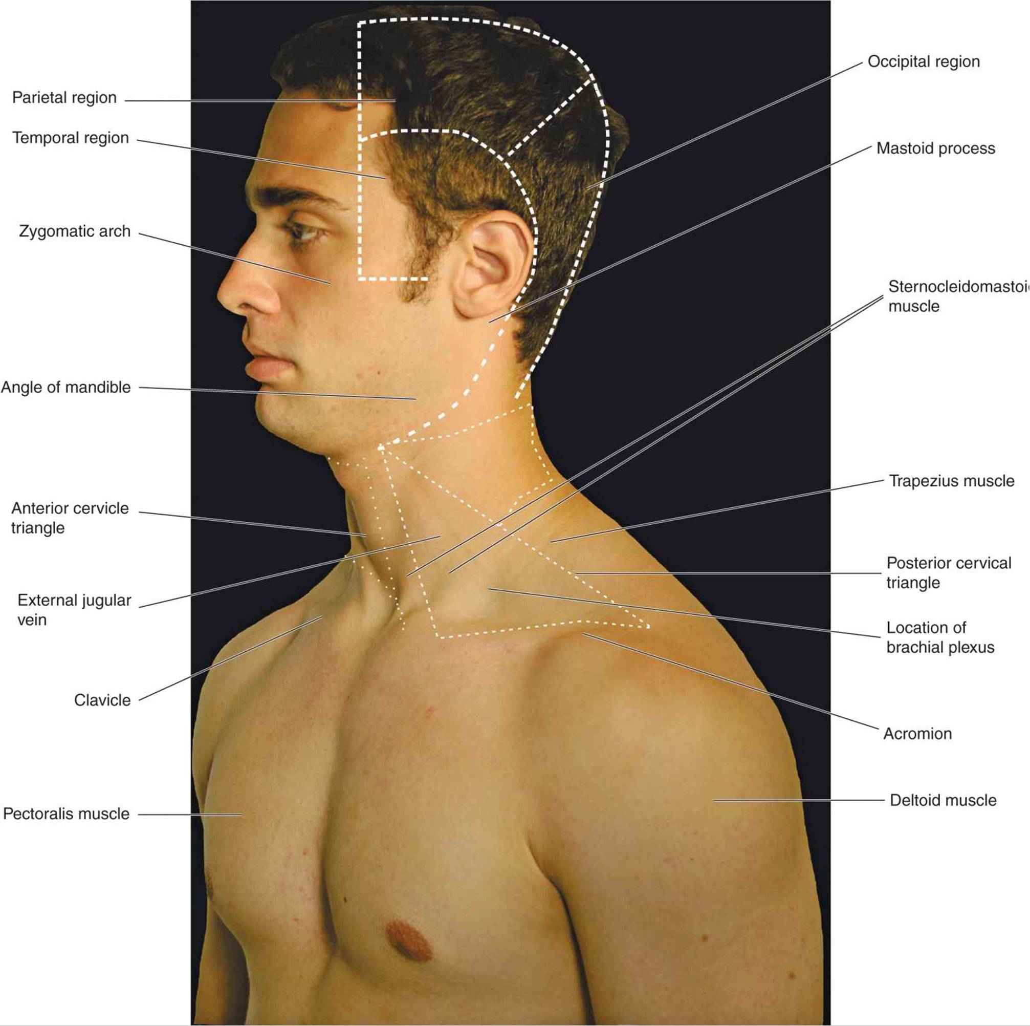
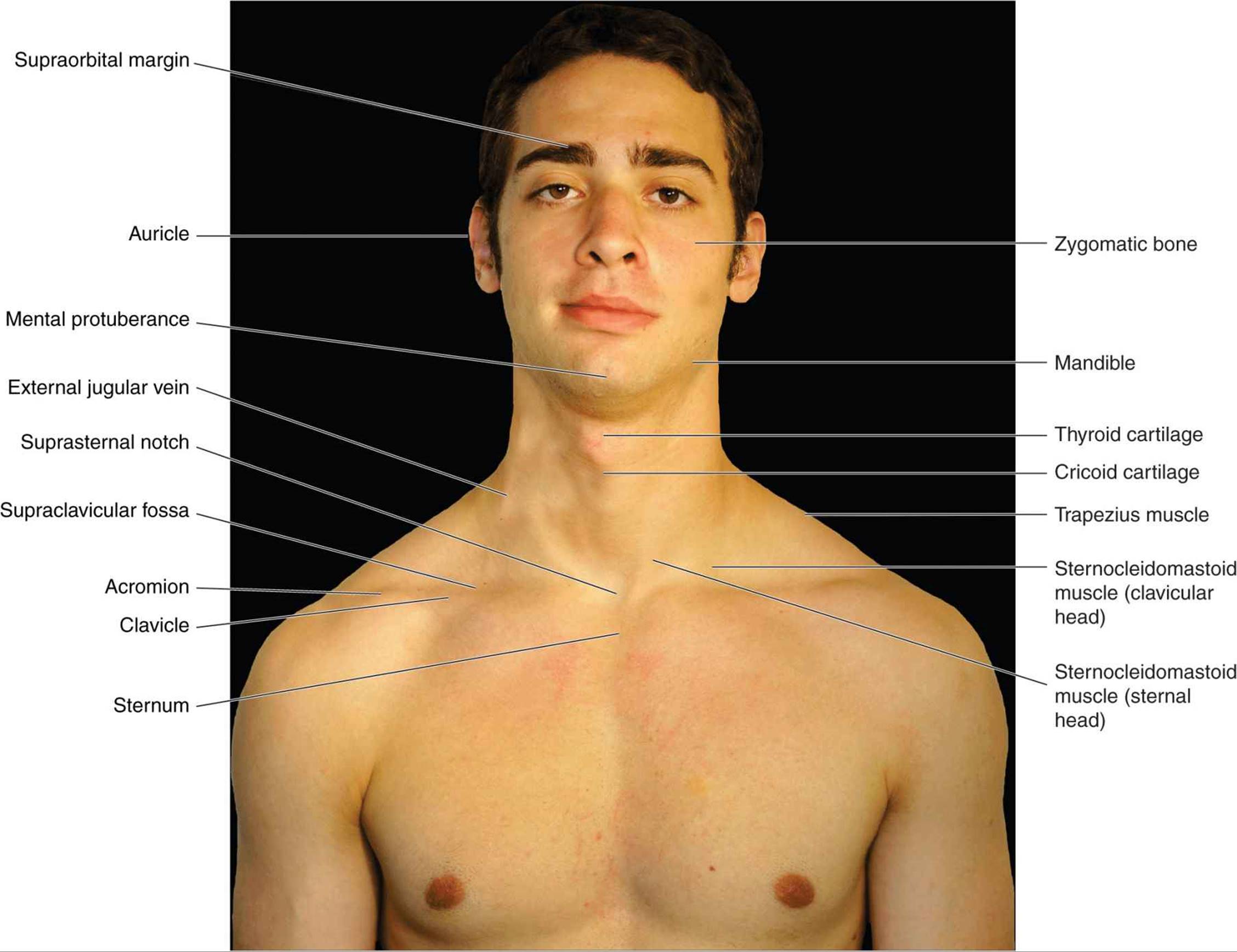
Anatomy Of The Upper Chest Area: Surface anatomy of anterior chest wall, spiral ct of thoracic inlet and surface anatomy of posterior chest wall.
Refference: Anatomy Of The Upper Chest Area